In the field of medicine, radiomics is a method that extracts large amount of features from radiographic medical images using data-characterisation algorithms. Radiomic features have the potential to uncover disease characteristics that fail to be appreciated by the naked eye. The hypothesis of radiomics is that the distinctive imaging features between disease forms may be useful for predicting prognosis and therapeutic response for various conditions, thus providing valuable information for personalized therapy. Radiomics emerged from the medical field of oncology and is the most advanced in applications within that field. However, the technique can be applied to any medical study where a disease or a condition can be imaged.
The imaging lab works on the investigation of textural features of several tumours as a potential additional pre-surgical tool of lesion discrimination using a radiomic approach. Currently, our interests are focused on salivary gland, lung and breast tumours.
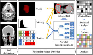
The underlying image data that is used to characterize tumors is provided by medical scanning technology. After the images have been saved in the database, they have to be reduced to the essential parts, in this case the tumors, which are called “volumes of interest”. After the segmentation, many features of the tumor may be computed. They stretch from volume, shape, surface to density and intensity as well as texture, tumor location, relations with the surrounding tissues and a lot of others.
After the selection of features that are important for our task it is crucial to analyze the chosen data.
Comments are closed