Senza categoria
Call for Research Grants 2024
Two research grants are available now and will expire on 20/07/2024 at 1 pm:
#1: https://bandi.unipi.it/
public/Bandi/Detail/cbc530e2- 8bc0-415f-9378-72031ea0bfeb #2: https://bandi.unipi.it/
public/Bandi/Detail/739b5a48- 57b5-4feb-8706-31580f14aa9c PRIMAGE project – Aiming AI at lethal paediatric tumours
La Fe University and Polytechnic Hospital in Valencia, Spain, is coordinating EU-funded program PRIMAGE, which uses precision information from medical imaging to advance knowledge of the most lethal paediatric tumours, by establishing their prognosis and expected treatment response using radiomics, imaging biomarkers and artificial intelligence (AI).
Read more on https://healthcare-in-europe.com/en/news/aiming-ai-at-lethal-paediatric-tumours.html
EUSOMII on AIR interview
13 Jan. 2021 – Interview of Prof. Emanuele Neri from EUSOMII
In this first 2021 episode of EuSoMII on Air, Shah Islam and Merel Huisman interview Prof. Emanuele Neri (University of Pisa), who is one of the co-founders of EuSoMII about the early days of EuSoMII and the way imaging informatics was introduced as a well-defined domain of expertise in the European landscape of radiology. Prof. Neri also tells us about the significance and value of the ongoing A.I. revolution that is taking place within the profession of radiology, and the importance of including basic knowledge about A.I. into the European Training Curriculum, for training the new generation of radiologists.
Radiomic analysis
In the field of medicine, radiomics is a method that extracts large amount of features from radiographic medical images using data-characterisation algorithms. Radiomic features have the potential to uncover disease characteristics that fail to be appreciated by the naked eye. The hypothesis of radiomics is that the distinctive imaging features between disease forms may be useful for predicting prognosis and therapeutic response for various conditions, thus providing valuable information for personalized therapy. Radiomics emerged from the medical field of oncology and is the most advanced in applications within that field. However, the technique can be applied to any medical study where a disease or a condition can be imaged.
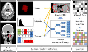
The imaging lab works on the investigation of textural features of several tumours as a potential additional pre-surgical tool of lesion discrimination using a radiomic approach. Currently, our interests are focused on salivary gland, lung and breast tumours.
The underlying image data that is used to characterize tumors is provided by medical scanning technology. After the images have been saved in the database, they have to be reduced to the essential parts, in this case the tumors, which are called “volumes of interest”. After the segmentation, many features of the tumor may be computed. They stretch from volume, shape, surface to density and intensity as well as texture, tumor location, relations with the surrounding tissues and a lot of others.
After the selection of features that are important for our task it is crucial to analyze the chosen data.
Artificial Intelligence
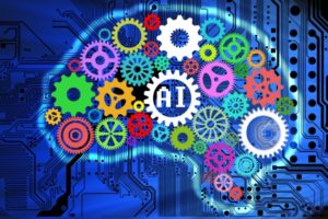
In computer science, artificial intelligence (AI), sometimes called machine intelligence, is intelligence demonstrated by machines, in contrast to the natural intelligence displayed by humans. Leading AI textbooks define the field as the study of “intelligent agents”: any device that perceives its environment and takes actions that maximize its chance of successfully achieving its goals. Colloquially, the term “artificial intelligence” is often used to describe machines (or computers) that mimic “cognitive” functions that humans associate with the human mind, such as “learning” and “problem solving”.
AI in healthcare is often used for classification, whether to automate initial evaluation of a CT scan or EKG or to identify high risk patients for population health. The breadth of applications is rapidly increasing. As an example, AI is being applied to the high cost problem of dosage issues—where findings suggested that AI could save $16 billion. In 2016, a ground breaking study in California found that a mathematical formula developed with the help of AI correctly determined the accurate dose of immunosuppressant drugs to give to organ patients.
Artificial intelligence is assisting doctors. There is a great amount of research and drugs developed relating to cancer. Our work focuses on classification of specific pathological lesions as a potential additional non-invasive pre-surgical tool.